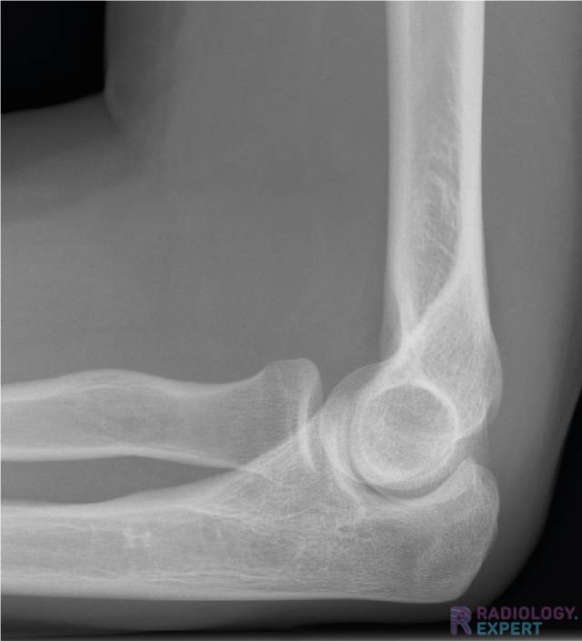
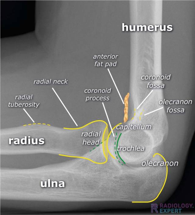
X-Elbow
The basic principles about the elbow X-ray examination.

X-ELBOW - Introduction
An X-ray of the elbow is a frequently conducted examination and is mainly used for diagnosing a fracture.
Some of the key topics are radial head fracture, supracondylar humeral fracture, anterior/posterior fat pad and elbow luxation.
Prior to this module, it is wise to read the Fracture General Principles module.
This module consists of the components indication/technique, normal anatomy, checklist and pathology.
KEY TOPICS/TERMS:
- Radial head fracture
- Radial head luxation
- Pulled elbow / Nursemaid's elbow
- Essex-Lopresti fracture
- Monteggia fracture
- Supracondylar humeral fracture
- Lateral humeral condylar fracture
- Medial humeral epicondylar fracture
- Olecranon fracture
- Elbow luxation
- Osteoarthritis


Interested?
Learn everything about X-Elbow
Text
drs. A. van der Plas (MSK radiologist Maastricht UMC+)
With special thanks to:
drs. M. Özdemir (radiologist Noordwest Ziekenhuisgroep)
dhr. A. Bubberman (Advanced Practioner Radiology LUMC)
Illustrations
drs. A. van der Plas (MSK radiologist Maastricht UMC+)
Sources:
- B.J. Manaster et al. The Requisites – Musculoskeletal Imaging (2007).
- N. Raby et al. Accident & Emergency Radiology – A Survival Guide. (2005).
- K.L. Bontrager, J.P. Lampignano. Textbook of Radiographic Positioning and Related Anatomy. 2014 (8th edition)
14/05/2016
(All the work (text, illustrations, visual elements) seen on this website is copyright by Radiology Expert.
It may not be used without written permission of Radiology Expert).
