Abdominal ultrasound
The basic principles of the abdominal ultrasound examination.

- Indication / Technique
- Normal Anatomy Liver & Gallbladder
- Normal Anatomy Urinary tract
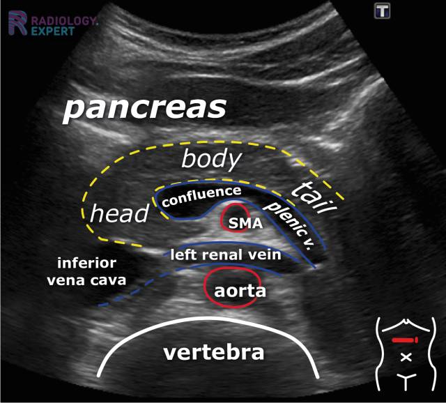
- Normal Anatomy Spleen & Pancreas
- Normal Anatomy Aorta
- Normal Anatomy Intestines
- Checklist
- Pathology Liver & Gallbladder
- Pathology Urinary tract
- Pathology Spleen & Pancreas
- Pathology Aorta
- Pathology Intestines & Trauma
Abdominal Ultrasound - Introduction
An abdominal ultrasound examination is essential in clinical practice. This examination allows for the assessment of various possibilities and limitations, as well as the frequent pathologies encountered, such as bile stones, steatosis, hydronephrosis, and appendicitis.
Prior to this module, it is wise to read the Ultrasound Technique module.
This module consists of the components indication/technique, normal anatomy, checklist and pathology (the organs are divided over multiple chapters).
KEY TOPICS/TERMS:
- Liver lesions
- Steatosis
- Bile stones
- Dilated bile ducts
- Kidney lesions
- Kidney stones
- Hydronephrosis
- Bladder tumor
- Splenomegaly
- Abdominal aneurysm
- Appendicitis
- Diverticulitis
- Intestinal wall thickening
- Free abdominal fluid

Interested?
Learn everything about Abdominal ultrasound
Text
drs. A. van der Plas (MSK radiologist Maastricht UMC+)
drs. M.P.M. Kop (abdominal radiologist Amsterdam UMC)
Illustrations
drs. A. van der Plas (MSK radiologist Maastricht UMC+)
Sources:
- B. Block. Abdominal Ultrasound: Step by Step (2004).
- W.D. Middleton et al. The Requisites – Ultrasound (2004).
30/07/2016
(All the work (text, illustrations, visual elements) seen on this website is copyright by Radiology Expert.
It may not be used without written permission of Radiology Expert).